దస్త్రం:Bronchiolar epithelium 3 - SEM.jpg
Jump to navigation
Jump to search

ఈ మునుజూపు పరిమాణం: 585 × 599 పిక్సెళ్ళు. ఇతర రిజల్యూషన్లు: 234 × 240 పిక్సెళ్ళు | 469 × 480 పిక్సెళ్ళు | 750 × 768 పిక్సెళ్ళు | 1,024 × 1,049 పిక్సెళ్ళు.
అసలు దస్త్రం (1,024 × 1,049 పిక్సెళ్ళు, ఫైలు పరిమాణం: 375 KB, MIME రకం: image/jpeg)
This is a file from the Wikimedia Commons. Information from its description page there is shown below. Commons is a freely licensed media file repository. You can help. |
సారాంశం
| వివరణ |
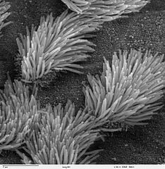
Scanning electron microscope image of lung trachea epithelium. There are both ciliated and non-ciliated cells in this epithelium. Note the difference in size between the cilia and the microvilli (on the non-ciliated cell surface). Zeiss DSM 962 SEM |
| మూలం | |
| కర్త | Charles Daghlian |
| అనుమతి (ఈ దస్త్రాన్ని పునర్వినియోగించుకోవడం) |
PD |
లైసెన్సింగ్
| ఈ కృతిని కృతికర్త, Charles Daghlian', సార్వజనీనంగా విడుదల చేసారు. ఇది ప్రపంచవ్యాప్తంగా వర్తిస్తుంది. కొన్ని దేశాల్లో ఇది చట్టబద్ధంగా సాధ్యంకాకపోవచ్చు; అయితే: ఈ కృతిని ఎటువంటి షరతులు లేకుండా, అట్టి షరతులు చట్టం వల్ల తప్పనిసరి అయితే తప్ప, ఏ ఉద్దేశానికైనా ఉపయోగించుకునే హక్కును ఎవరికైనా Charles Daghlian ప్రదానం చేస్తున్నారు.
|
ఫైలు చరితం
తేదీ/సమయం ను నొక్కి ఆ సమయాన ఫైలు ఎలా ఉండేదో చూడవచ్చు.
| తేదీ/సమయం | నఖచిత్రం | కొలతలు | వాడుకరి | వ్యాఖ్య | |
|---|---|---|---|---|---|
| ప్రస్తుత | 14:16, 7 అక్టోబరు 2006 | | 1,024 × 1,049 (375 KB) | Patho | {{Information |Description=Scanning electron microscope image of lung trachea epithelium. There are both ciliated and on-ciliated cells in this epithelium. Note the difference in size between the cilia and the microvilli(on non-ciliated cell surface) Zei |
లింకులు
కింది పేజీలలో ఈ ఫైలుకు లింకులు ఉన్నాయి:
సార్వత్రిక ఫైలు వాడుక
ఈ దస్త్రాన్ని ఈ క్రింది ఇతర వికీలు ఉపయోగిస్తున్నాయి:
- ar.wikipedia.org లో వాడుక
- ast.wikipedia.org లో వాడుక
- bs.wikipedia.org లో వాడుక
- ca.wikipedia.org లో వాడుక
- cs.wikipedia.org లో వాడుక
- da.wikipedia.org లో వాడుక
- de.wikipedia.org లో వాడుక
- de.wikibooks.org లో వాడుక
- en.wikipedia.org లో వాడుక
- es.wikipedia.org లో వాడుక
- eu.wikipedia.org లో వాడుక
- fa.wikipedia.org లో వాడుక
- fr.wikipedia.org లో వాడుక
- gl.wikipedia.org లో వాడుక
- he.wikipedia.org లో వాడుక
- he.wiktionary.org లో వాడుక
- hi.wikipedia.org లో వాడుక
- id.wikipedia.org లో వాడుక
- jv.wikipedia.org లో వాడుక
- kk.wikipedia.org లో వాడుక
- lt.wikipedia.org లో వాడుక
- lv.wikipedia.org లో వాడుక
- ms.wikipedia.org లో వాడుక
- nl.wikipedia.org లో వాడుక
- nn.wikipedia.org లో వాడుక
- no.wikipedia.org లో వాడుక
- pl.wikipedia.org లో వాడుక
- pl.wiktionary.org లో వాడుక
- pt.wikipedia.org లో వాడుక
- ru.wikipedia.org లో వాడుక
- ru.wiktionary.org లో వాడుక
- sh.wikipedia.org లో వాడుక
ఈ దస్త్రపు మరింత సార్వత్రిక వాడుకను చూడండి.